
Different shades of red revisited
January 29, 2008 § Leave a comment

A while back I wrote this post about the problems video cameras have with reproducing the insides of our bodies. The blood running through our bodies cause most tissues to be some shade of red.
This week I got an excellent opportunity to visualize this. I am currently filming a lot of operations for sarcomas of the upper and lower extremities. Depending on the situation of the tumor and other factors, the surgeons sometimes prefer to empty the blood from the surgical site before operating. An arm or leg can typically stay bloodless for about 2 hours before opening the tourniquet and letting the blood back in.
This technique allows the surgeons to work faster and more precisely as the separations of the different tissues and anatomical structures becomes more visible. The same applies to the camera. Without blood the highly saturated reds are gone, and you get colors that are easier to reproduce.
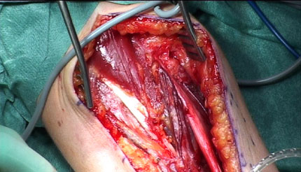
The video stills in this post are both from the same operation, an excision of a synovial sarcoma of the elbow. MRI indicated that the tumor infiltrated both major blood vessels and the median nerve. If this was the case, the arm could have to be amputated above the elbow. But the surgeons wanted to try to the excise the tumor first. Luckily there was hardly any infiltration at all and they were able to remove the tumor without damaging the surrounding structures.
The still above was taken just after the removal of the tumor. The median nerve can be seen going behind the scissors. The arm is still empty of blood at this stage and the blood vessels, nerves, muscles and fat are easily separated.
The still below was taken a few minutes after opening of the tourniquet. As you can see, all tissues now have a reddish color, making it harder to distinguish between them.

Leave a comment