
Sarcoma videos
June 20, 2008 § 13 Comments

Today, seven videos I’ve made of diagnosis and treatment of sarcomas were published on www.oncolex.org. Working on these videos has been especially interesting, as sarcomas are a very rare form of cancer, and because of all the devoted specialists I’ve gotten to know.
The hospital where I work is the national sarcoma center, and all sarcoma patients in Norway sent there. All evaluation and decisions on treatment is done by a team which includes radiologists, pathologists, oncologists, orthopedic surgeons, gastrointestinal surgeons and gynecologists. Other specialists, such as head and neck surgeons and thoracic surgeons, are consulted when needed. A very efficient way to work which ensures the best possible treatment for all patients. In Norway about 200 people develop some form of sarcoma every year (160 soft tissue sarcomas, 40 bone sarcomas). That’s only about 1 % of all malignant tumors diagnosed yearly.
In addition to footage of the diagnostic procedures and operations, all videos includes x-rays, MRI and/or CT images to visualize the tumor and its relation to anatomical structures.
Ultrasound-guided core needle biopsy
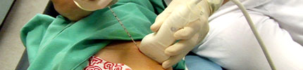 A diagnostic procedure performed to establish the histopathologic diagnosis of a soft tissue tumor on the right flank of a young teenage girl. The radiologist uses ultrasound to guide the insertion of the core needle. I don’t know the result. After the biopsy you can see that the radiologist tattoos the area where he inserted the needle. As it can be contaminated with tumor cells, the surgeons will need to remove the biopsy channel if the tumor have to be surgically removed.

CT-guided bone biopsy
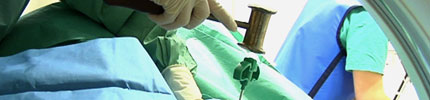 Also an image-guided diagnostic procedure. CT is used at intervals during the procedure to guide the insertion of a bone biopsy needle, in this case into a tumor of the left tibia of a young man. Both to establish if the tumor is benign or malignant and its histopathologic subtype.
I’ve written about the filming of this procedure before.
Wide excision of a liposarcoma of the thigh
 A video of an operation to remove a liposarcoma located in the vastus medialis muscle of the left thigh. The tumor have been biopsied prior to the operation. An eye-shaped incision is made around the biopsy channel and it is included in the surgical specimen. During the operation the femoral vein and artery and the saphenous nerve are exposed and dissected. The vastus medialis is cut at both ends, and the entire muscle is removed with the tumor left unexposed inside.
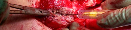
Excision of a chondrosarcoma of the humerus
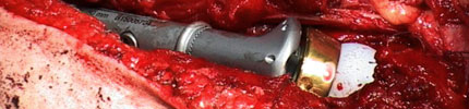 This was a patient with a large chondrosarcoma in the proximal end of the left humerus. Here too the biopsy channel is identified and included in the specimen. The different muscles covering the humerus are cut and the radial nerve is identified and dissected. The bone is cut about 5 cm below the tumor and the shoulder joint is opened.
The joint and proximal humerus is then reconstructed using a titanium prosthesis. I don’t remember why, but the joint was reversed with the cup on the humerus part of the prosthesis and the ball on the scapula.
It was a great challenge for me to try to identify all the muscles of the upper arm when editing. I expected I’d done a lot of mistakes, but on viewing the video, the surgeon found none! I guess I’ve learnt a thing or two along the way after all.
Resection of a sarcoma of the uterus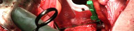 A radical hysterectomy to remove a leiomyosarcoma of the uterus. Performed as a normal hysterectomy except for the division of the vagina below the cervix. To avoid contaminating the pelvis and lower vagina with tumor cells, two rows of staples are placed on the vagina and it is then divided between them.
This is actually one of the first videos I made for this project, some three years ago, but not published until now.
Resection of a gastrointestinal stromal tumor (GIST)
 An operation to remove a large GIST of the stomach. The tumor originated in the cranial part of the stomach and grew laterally to the left, adhering to the spleen and the tail of the pancreas. There’s a characteristic fistula from the lumen of the stomach into the tumor. The tumor is dissected circumferentially. The spleen is included in the specimen. The pancreas tail is divided from the pancreas, using a TA stapler and knife, and also included.
I’ve written about the smell of this operation before.
Resection of a retroperitoneal sarcoma
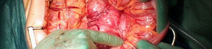 A large (⌀≈15 cm) tumor located in the left upper quadrant of the abdomen. The tumor grows through the mesocolon and shows a naked surface against the peritoneum. It’s close to the left kidney and pancreas but not adherent to any of them. Circumferential dissection of the tumor. Because the blood supply to the descending colon is involved in the tumor a left hemicolectomy is performed and the colon segment is included in the surgical specimen.
When the tumor is removed, the renal vein, splenic vein and abdominal aorta are exposed.
This video is currently not available.
The picture at the top of the post is a microscopic image of a stained fine needle aspiration from a chordoma.

Really nice work! I watched most of the videos and will check back later to see the others.
Thanks!
Great videos! I’m a pediatric oncologist and I run the musculoskeletal tumor program at my hospital, so I especially was interested in watching these videos. It was fun trying to translate the Norwegian into English in my head. I haven’t had time to watch them all, but I can’t wait to see the ones I’ve missed.
Thanks, David!
I didn’t film any operations on children this time around, but working on sarcomas I heard a lot of stories both sad and encouraging about children with (mostly osteo-) sarcomas.
One operation I have not gotten the opportunity to film yet is “rotation-plasty”. What a strange, but great idea!
You MUST let me know if you ever do film a rotationplasty. I’ve taken care of kids after one of those, but have never seen it done in the OR.
I will!
I’m also going to film a sacrectomy, whenever one of those are scheduled.
good stuff!
I really appreciate that you have a video of a humerus chondrosarcoma. It will prove helpful for those who have such a diagnosis to understand better what it is their surgeons have done to save their limb! Do you have the opportunity to go to other countries to film surgeries?
Thank you, Elizabliss. The possibilities of limb sparing surgery is truly amazing.
As I am employed by a hospital here in Norway, filming abroad is basically not easy to do. But please contact me if you have any specific proposals. I’ll be more than happy to discuss it.
Thanks for posting these videos. I have had 8 retroperitoneal liposarcoma surgeries and I always wish that I could leave my video camera on during the surgery…. I am glad I found your blog :))
Thank you, Elsa! That means a lot to me. I hope your doing fine.
[…] of the first operations I ever filmed is a good example. It was a hysterectomy to remove a uterine sarcoma. A gynoncologist on my project was going to operate, and we were OK to go. I went to the OR, rigged […]
that was amazing. It was very interesting. all though i couldnt understand it all. i still picked up on some of the meaning.